What Is the KLOW Blend?
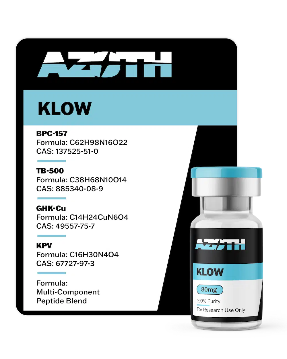
The KLOW Blend is a four-component peptide formulation combining BPC-157, TB-500 (full 43 amino acid Thymosin Beta-4), KPV, and GHK-Cu into a single lyophilized research preparation. Supplied in a standardized 80mg format, the blend is designed for qualified laboratory research and analytical investigation programs requiring access to multiple peptide research compounds within a unified formulation.
Each compound in the KLOW Blend has been independently studied across distinct preclinical research areas. BPC-157, a 15 amino acid gastric pentadecapeptide, has been characterized for its effects on nitric oxide signaling and vascular endothelial biology in animal models. TB-500, the full 43 amino acid sequence of Thymosin Beta-4, has been studied for its role in cytoskeletal actin regulation and cell motility in preclinical systems. KPV, the C-terminal tripeptide fragment of alpha-MSH, has been investigated for modulation of NF-kappaB and melanocortin immune signaling pathways. GHK-Cu, a naturally occurring copper-binding peptide complex, has been characterized for effects on metalloproteinase activity, gene expression modulation, and oxidative stress regulation in in vitro and animal model research contexts.
Together, these four compounds span a range of cellular signaling pathways relevant to vascular biology, extracellular matrix research, inflammatory pathway analysis, and cytoskeletal dynamics. The KLOW Blend provides researchers with a multi-compound formulation for evaluating peptide behavior, pathway-selective interactions, blend stability, and analytical performance across related peptide systems within controlled laboratory research programs. This product is supplied strictly for laboratory and research use only and is not intended for human or veterinary use, diagnostic use, or any therapeutic application.
RESEARCH USE ONLY: NOT FOR HUMAN OR VETERINARY USE
KLOW Blend Specifications
| Component | Dose | CAS | Molecular Formula | MW (g/mol) | Purity |
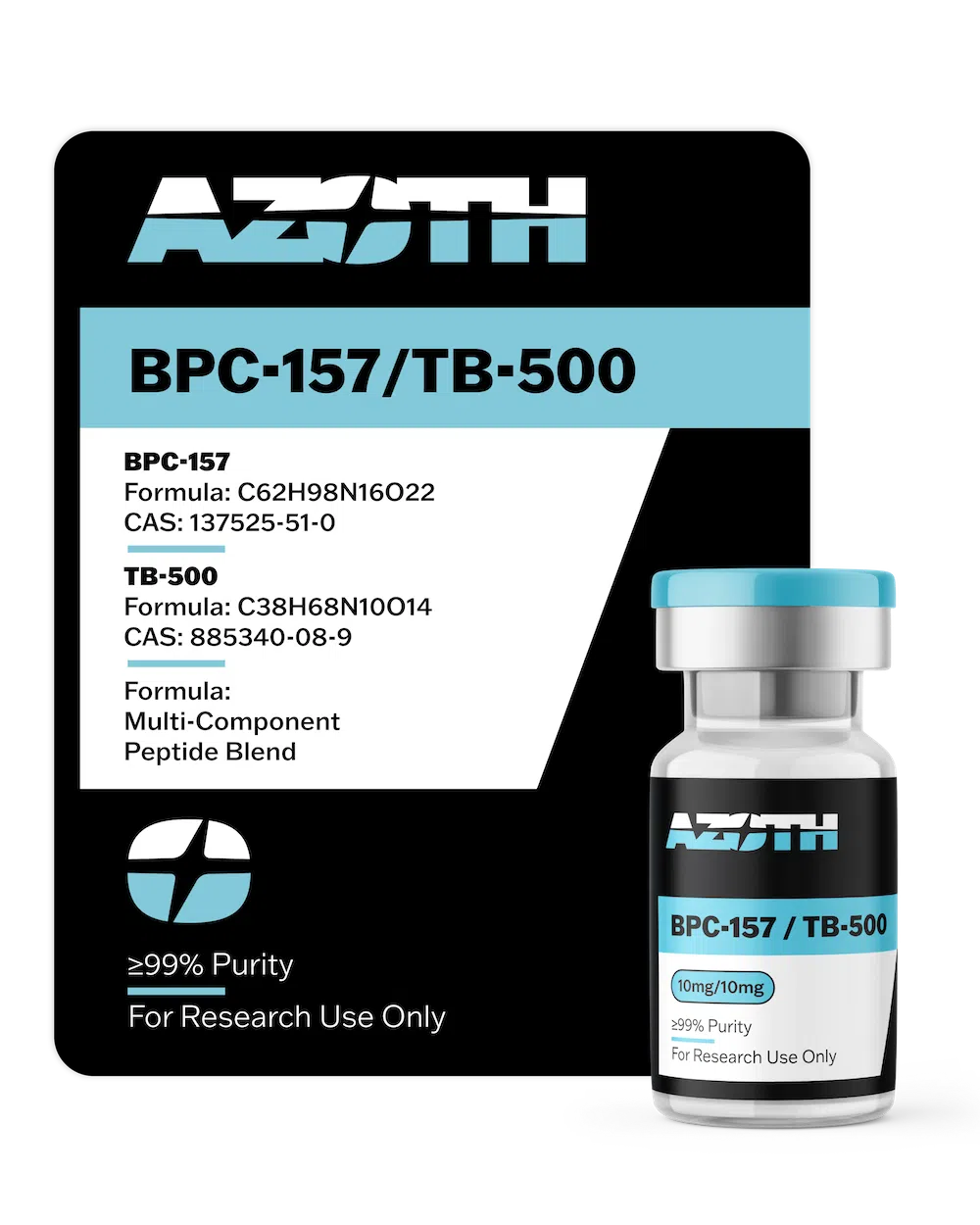
| BPC-157 | 10mg | 137525-51-0 | C62H98N16O22 | 1419.556 | 99% |
| TB-500 (Thymosin Beta-4) | 10mg | 77591-33-4 | C212H350N56O78S | 4963.44 | 99% |
| KPV | 10mg | 112965-21-6 | C17H32N6O4 | 384.48 | 99% |
| GHK-Cu | 50mg | 89030-95-5 | C14H23CuN6O4 | 401.91 | 99% |
| Total Blend | 80mg |
BPC-157: Molecular Profile and Preclinical Research Background
BPC-157 is a synthetic 15 amino acid pentadecapeptide derived from a sequence found in a naturally occurring protein in human gastric juice. Designated Body Protection Compound-157, this stable gastric pentadecapeptide has been studied extensively in preclinical model systems spanning gastroenterology, vascular biology, musculoskeletal research, and cellular regeneration science. Animal model research, conducted predominantly in rodents, has documented the compound’s observed effects across gastrointestinal mucosa, tendon, ligament, skeletal muscle, and vascular endothelium in non-clinical settings.
BPC-157 appears to operate through several interconnected signaling cascades rather than a single receptor pathway, with preclinical data characterizing it as a pleiotropic regulatory compound. Among the most consistently documented preclinical findings is the compound’s modulation of the nitric oxide system. Studies have shown that BPC-157 promotes the phosphorylation of Src, Cav-1, and eNOS while reducing the Cav-1 and eNOS binding interaction, a regulatory step in eNOS activation and NO production in vascular model systems. This eNOS modulation is associated with several of the compound’s observed effects on vascular endothelial cell migration and blood vessel formation in animal models.
Preclinical data have also documented BPC-157’s upregulation of Vascular Endothelial Growth Factor Receptor 2 (VEGFR2), sensitizing endothelial cells to circulating growth factors through the VEGFR2-Akt-eNOS signaling axis. Rodent ischemia and injury model studies have documented enhanced collateral blood vessel formation following administration. In fibroblast model systems, BPC-157 stimulates the FAK-paxillin pathway, influencing cellular adhesion and migration relevant to extracellular matrix research and wound model studies, with dose-dependent effects documented in both in vitro and in vivo experimental contexts.
Gene expression analysis in preclinical research has documented BPC-157’s modulation of Egr, Nos (particularly eNOS), Srf, Vegfr, Plcgamma, and Kras, regulatory genes with influence across vascular and immune cell behavior. Expression effects on each gene vary with time following administration, indicating a time-dependent regulatory mechanism with broad control over multiple target genes. Pharmacokinetic studies in rats and dogs have documented systemic distribution within approximately 10 minutes of administration, with peak tissue concentrations in the kidney, liver, thymus, and spleen observed at approximately one hour post-administration.
TB-500: Molecular Profile and Preclinical Research Background
TB-500 is a synthetic peptide corresponding to the full 43 amino acid sequence of Thymosin Beta-4 (Tbeta4), a naturally occurring protein found in virtually all nucleated cells of the body. Thymosin Beta-4 is among the most abundant intracellular proteins in mammalian tissue and has been detected at elevated concentrations in platelets and wound fluid. Preclinical research on TB-500 spans cardiovascular regeneration biology, musculoskeletal repair models, ocular surface research, and immune cell trafficking studies in rodents and in vitro systems.
TB-500 operates through two characterized mechanisms in the preclinical literature. The first involves actin sequestration: the compound binds to G-actin monomers, regulating the pool of unpolymerized actin available for cytoskeletal assembly. This interaction influences cell motility, morphology, and division, and has been studied in immune cell migration, endothelial sprouting, and tissue repair model systems in vitro and in rodents.
The second mechanism, described in the research literature as a “moonlighting” function, involves gene expression modulation operating independently of actin binding. Through this pathway, TB-500 has been shown to influence the expression of genes involved in NO production, angiogenic signaling, and cellular proliferation. It also downregulates NF-kappaB and Toll-like receptor signaling, reducing the expression of pro-inflammatory cytokines including TNF-alpha and IL-1 receptor-associated kinases in preclinical model systems, with several downstream effects paralleling those observed with BPC-157 in comparable animal model contexts.
TB-500 activates additional tissue repair signaling cascades documented in preclinical data, including PI3K/Akt/eNOS, Notch, and angiopoietin-1/Tie pathways. Its modulation of the TGF-beta pathway has been studied in the context of fibrosis-related and connective tissue repair research in animal models. In cardiovascular biology studies, TB-500 has been shown to promote epicardial progenitor cell activation and myocyte survival following experimental cardiac injury in animal models. Phase 2 clinical evaluation of thymosin beta-4 has been conducted in chronic skin wound model settings including pressure ulcers and epidermolysis bullosa, where the compound was assessed for safety and tolerability.
KPV: Molecular Profile and Preclinical Research Background
KPV is a tripeptide composed of the sequence Lys-Pro-Val, representing the C-terminal tripeptide fragment of alpha-melanocyte-stimulating hormone (alpha-MSH). Despite its three amino acid structure and molecular weight of approximately 384 g/mol, KPV has accumulated a body of preclinical research characterizing its immune signaling modulation and inflammatory pathway activity. Research programs have examined KPV’s effects in gastrointestinal model systems, pulmonary research models, and central nervous system inflammation studies conducted in rodents.
The compound’s anti-inflammatory profile was first characterized in the mid-1980s, when animal model studies documented activity across experimental models of fever, dermatitis, vasculitis, fibrosis, arthritis, uveitis, gastritis, and inflammatory conditions affecting the lungs and central nervous system. Subsequent mechanistic research identified multiple pathways through which KPV modulates inflammatory signaling in preclinical systems: inhibition of NF-kappaB, downregulation of adhesion molecule and chemokine receptor expression, suppression of pro-inflammatory cytokine production, regulation of T-cell activity and inflammatory cell migration, and modulation of apoptosis-related signaling pathways.
While KPV interacts with melanocortin receptors and demonstrates binding affinity for MC1R, preclinical evidence indicates that its anti-inflammatory effects operate substantially independent of melanocortin receptor binding, distinguishing its mechanistic profile from the parent molecule alpha-MSH. Animal model studies have documented a tissue-selective pattern of activity, with KPV’s anti-inflammatory effects appearing concentrated in tissues characterized by excessive or pathological inflammation while leaving physiologically normal inflammatory processes largely unaffected.
Stability research has shown that unmodified KPV degrades to its constituent amino acids within approximately 24 hours under simulated physiological conditions, while glycoalkylation modifications extend stability without reducing biological activity in vitro. KPV has also been characterized for antimicrobial activity in laboratory settings, with documented effects against Staphylococcus aureus and Candida albicans, associated in part with cellular cAMP elevation and neutrophil response modulation.
GHK-Cu: Molecular Profile and Preclinical Research Background
GHK-Cu is a naturally occurring copper complex consisting of the tripeptide glycyl-l-histidyl-l-lysine (GHK) bound to a copper(II) ion. Initially isolated from human plasma and subsequently detected in saliva and urine, GHK-Cu is one of the most extensively characterized copper-peptide complexes in preclinical and in vitro research. The compound’s research profile spans extracellular matrix remodeling studies, gene expression analysis, oxidative stress regulation, pulmonary biology models, and connective tissue research systems.
GHK-Cu’s best-characterized mechanism involves regulation of metalloproteinase enzyme activity. The compound stimulates the production of matrix metalloproteinases, enzymes responsible for breaking down damaged extracellular matrix proteins during tissue remodeling, while simultaneously promoting anti-protease expression to preserve intact structural proteins from degradation. This coordinated regulation of protein synthesis and breakdown has been documented in skin and connective tissue model systems and is associated with GHK-Cu’s observed effects on extracellular matrix organization in preclinical wound model research.
GHK-Cu has been characterized for broad effects on gene expression in in vitro systems. An analysis of its gene expression influence reported modulation of activity across approximately 32% of human genes, including suppression of genes associated with inflammatory signaling and tissue degradation, and promotion of genes associated with cellular repair pathways. Additional mechanistic data document GHK-Cu’s suppression of pro-inflammatory cytokines including TNF-alpha and IL-6, inhibition of NF-kappaB activity, neutralization of reactive oxygen species (ROS), and enhancement of antioxidant enzyme production including superoxide dismutase and glutathione in in vitro and animal model systems.
Pulmonary research in rodent models has documented GHK-Cu’s effects on lung inflammation and oxidative stress parameters in LPS-challenge and cigarette smoke exposure studies. Neurological model research has examined GHK-Cu’s influence on copper homeostasis and amyloid beta aggregation, areas of interest in neurodegenerative disease research programs. In laboratory settings, GHK-Cu has also been characterized for antimicrobial activity, with evidence of antimicrobial complex formation via interaction with fatty acids released from damaged tissue in vitro.